Clinical Cases to Challenge, Teach, and Refine Your Ophthalmic Practice
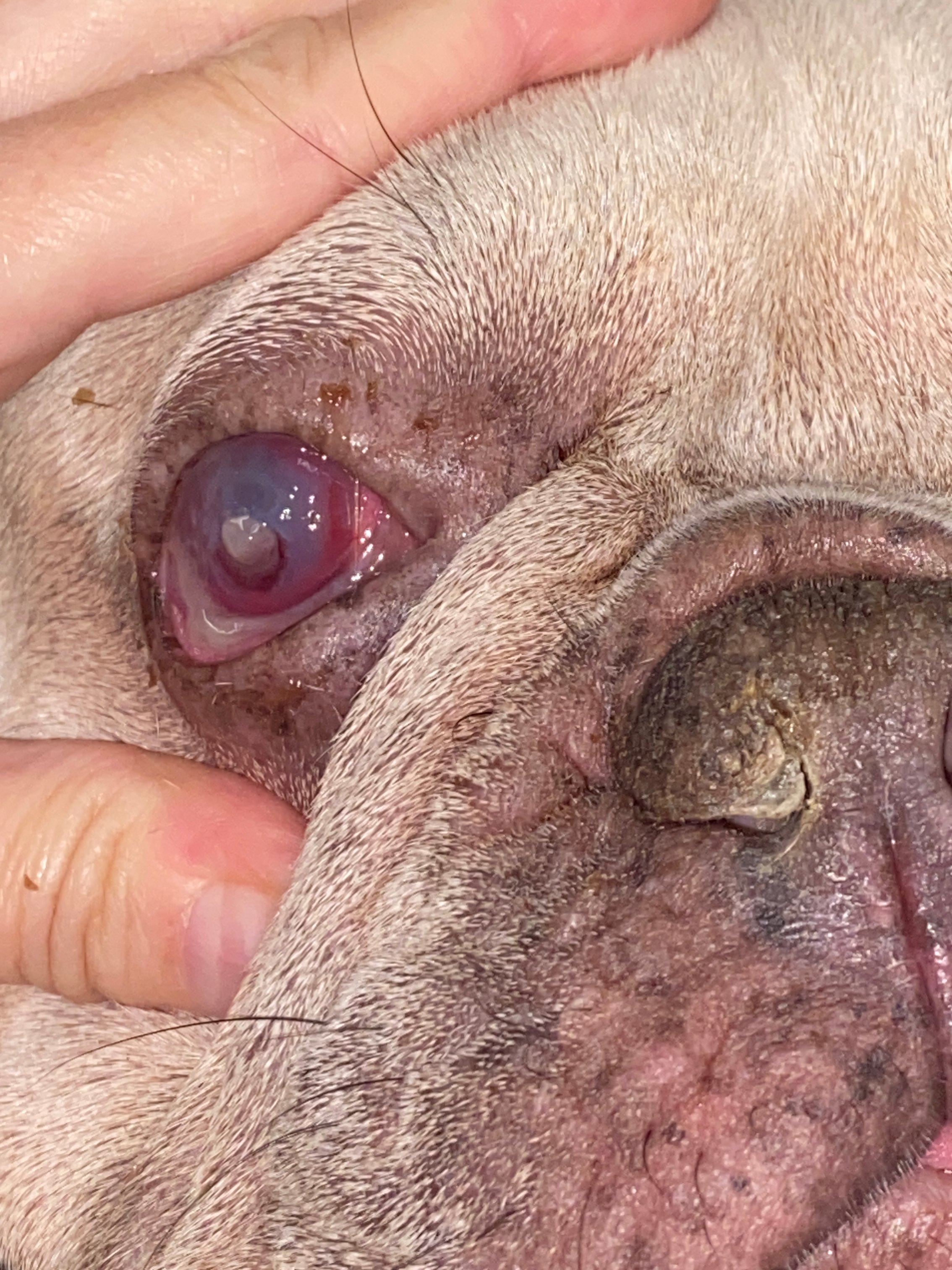
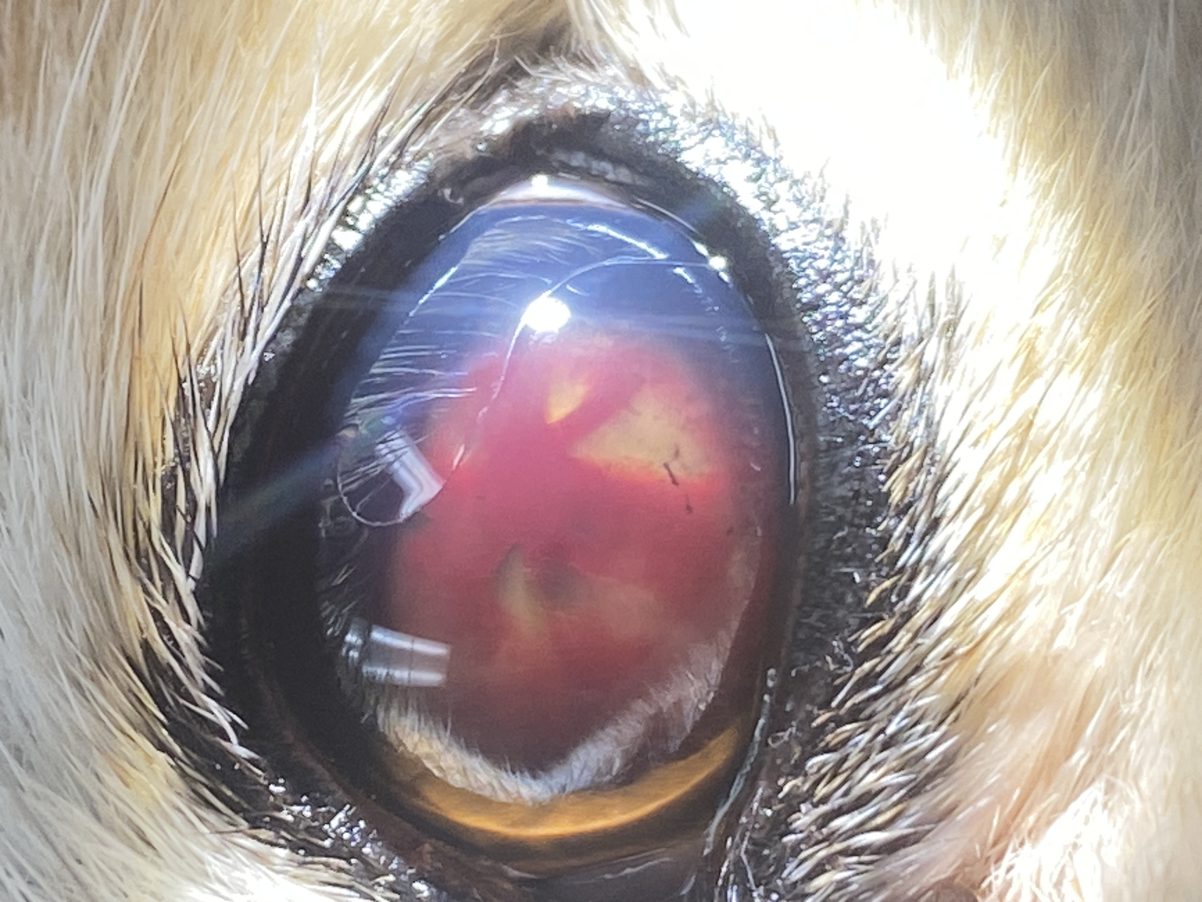
Welcome to a curated collection of real-world ophthalmic cases drawn from clinical practice. Each image is paired with a focused clinical insight and a key takeaway designed to sharpen your diagnostic thinking and refine your treatment approach. From corneal ulcers and hypopyon to uveal cysts, hyphema, and beyond, these cases reflect the complexity and nuance that defines veterinary ophthalmology in practice.
Whether you're a seasoned clinician or still building your ophthalmic confidence, this page is designed to challenge assumptions, highlight the findings that matter most, and prompt the kind of critical thinking that changes outcomes. Each case invites you to look closer, consider the differential, and ask what you might have missed.
Scroll through, engage with the questions, and test your clinical instincts — because in ophthalmology, what you see is only the beginning.
Clinical Insight:
Corneal edema with vascularization and a hypopyon can be seen in both severe uveitis and infectious corneal disease. The key distinction is whether the cornea itself is compromised. Subtle or delayed fluorescein uptake, focal opacity, and marked pain should raise suspicion for a deep ulcer or early abscess. Look at the circular yellow infiltrate nasally at the terminus of the vessels and hypopyon
Takeaway:
If there’s any doubt, treat as an ulcer first. Missing an infection and reaching for steroids or even a topical NSAID can rapidly worsen the outcome and potentially worsen the visual prognosis.
Clinical Insight:
The fluorescein highlights a superficial epithelial defect, but the adjacent darker facet represents deeper stromal loss that does not stain. The surrounding 360° vascularization signals an active, chronic healing response—and a cornea under significant stress.
Takeaway:
Not all ulcers are created equal. Identifying deeper stromal involvement changes both urgency and treatment strategy.
Would you manage this medically, or refer for surgical support? Would you consider debridement?
Clinical Insight:
This is bullous keratopathy—fluid accumulation within the cornea leading to formation of epithelial bullae. In this case, the bulla is large enough to protrude beyond the normal corneal contour, with surrounding vascularization indicating chronicity and stress.
Takeaway:
Even when fluorescein is negative, these bullae are fragile. Rupture can rapidly convert this into a painful ulcerative emergency.
At what point do you move from medical management to surgical intervention in cases like this? Do you know the single most important treatment modality in this case? Hint: It was named after the ruffled, high-collared garments worn by nobility in the 1500s.
Clinical Insight:
Severe conjunctival hyperemia and thickening like this are classic for feline herpesvirus-1 (FHV-1). Even when the cornea is initially intact, viral reactivation can lead to ulceration—especially if local immunity is suppressed.
Takeaway:
In suspected herpetic disease, avoid topical steroids early. Antiviral therapy and supportive care should come first.
What other diseases can mimic FHV 1 and produce conjunctivitis in cats?
Clinical Insight:
This dorsal superficial ulcer with focal vascularization is in a classic location for irritation from ectopic cilia—hairs emerging from the palpebral conjunctiva and contacting the cornea with every blink. These cases often fail to heal until the source of irritation is removed.
Takeaway:
Chronic or recurrent ulcers should always trigger a search for an underlying cause—especially eyelid abnormalities.
Do you routinely evert the eyelids in non-healing ulcers and look with magnification?
Clinical Insight:
This is a uveal cyst—typically arising from the iris or ciliary body and often free-floating in the anterior chamber. Most are incidental findings and don’t require treatment if the eye is otherwise normal, and the cyst is not interfering with vision.
Takeaway:
Differentiate cyst vs tumor: cysts are thin-walled, often mobile, and transilluminate—melanomas are solid and fixed (and associated with the iris).
In what breed could a uveal cyst trigger concern for something more sinister?
Clinical Insight:
Unlike a uveal cyst, this pigmented lesion is fixed to the iris (and is actually displacing the iris forward in this case) and causes distortion of the pupil (dyscoria). Solid, non-mobile, and progressive lesions raise concern for iris melanoma.
Takeaway:
Mobility matters. If it doesn’t move, don’t assume it’s benign.
When is it time to refer?
Bonus:
|
Feature |
Uveal Cyst |
Iris Melanoma |
|
Mobility |
Moves / floats |
Fixed |
|
Structure |
Thin-walled |
Solid |
|
Shape |
Smooth, spherical |
Irregular, raised |
|
Effect on pupil |
None |
Dyscoria |
|
Progression |
Stable |
Progressive |
|
Risk |
Usually benign |
Potentially serious |
Clinical Insight:
A central corneal perforation with a fibrin plug is surrounded by multiple stromal ulcers and corneal inflammation, reflecting a compromised and unstable ocular surface. In this case, severely reduced tear production led to chronic inflammation, poor healing, and eventual structural failure of the cornea.
Takeaway:
Chronic discharge isn’t benign—undiagnosed KCS can progress to vision-threatening complications, including perforation, especially in brachycephalic breeds with poor corneal sensation to start.
How early do you reach for a Schirmer tear test in chronic “goopy eye” cases? What does a dry nostril tell you about treatment prognosis in cases of KCS?

prognosis.
Clinical Insight:
Persistent hyperplastic primary vitreous (PHPV) is a developmental anomaly where fetal vasculature fails to regress, leaving a fibrovascular membrane behind the lens. These abnormal vessels can bleed, leading to hyphema and progressive intraocular changes, including cataract formation.
Takeaway:
When you see a young dog with cataract + hyphema, think congenital—not just inflammatory or traumatic causes.
Clinical Insight:
This hyphema is no longer acute—note the organized, fibrinous appearance. As blood breaks down and organizes within the anterior chamber, it can obscure deeper structures and complicate intraocular dynamics. The concurrent cataract further limits visualization and may be part of the underlying disease process.
Takeaway:
Hyphema requires investigation, not just treatment. Trauma, hypertension, coagulopathy, and intraocular disease should all be considered.
What ophthalmic diagnostic should ALWAYS be performed in these cases?